Farewells, Thank Yous, and Many Smiles

Greetings again!

     This post is a long time coming.  I couldn't end this blog without some type of farewell and thank you to all of the people who have helped me on this incredible journey.

First off - the presentation.  Thank you to everyone who came despite the fact it was a Saturday afternoon and the weather was bearable in terms of Tucson standards. I think I can speak for all of us who presented that day that it was so much fun sharing what we've been up to for the past few months.  For anyone who is curious...the link to my presentation is below:

https://app.emaze.com/@ALLRRCTF/srp

I don't have many words on the slides, but I might upload my presentation notes for anyone who wants to get a basic just of my project without reading through the long (and frankly quite tedious) posts on this blog.

     I am, of course, most grateful to Dr. Kasoff, my SRP mentor, for offering me (the incredibly inexperienced and unknowledgeable high liability high school student) this opportunity and seeing it through until the end.  His schedule was always full, yet he still willingly took the time to answer my questions, meet with me to discuss my project, and allow me to shadow him while he treated his patients.
     I originally began this project looking forward to OR time the most.  To me, it was the coolest thing, being able to observe operations in the flesh (no pun intended) rather than watching through a computer screen via YouTube.  Yet, after finishing this project, I found myself enjoying witnessing patient/doctor/nurse interactions more than anything.  It's very cliche and expected - when treating patients, you are exposed to the stories of all these strangers, every one of them has an enthralling tale to tell.  Cliche as it is, the stories are addicting, and you can't help but be captivated by each one.  The elderly couple, both with walking impairments, both accompanying each other into surgery.  The other couple in their mid sixties who still joke around like teenagers...
     So I can say my biggest lesson wasn't the procedures I learned or the results of the many research papers I memorized.  My biggest lesson was in patient care and bedside manners - lessons that cannot be taught by textbooks.  Watching Dr. Kasoff, the various nurses, residents, and technicians treat the patients has been my most significant takeaway.
     In fact, this entire project has broadened the scope of my understanding of the field of medicine.  The term "teaching hospital" has taken on a new meaning.  During the operation, various technicians, nurses, doctors, residents, and company representatives would approach me asking if I had any questions and enthusiastically answered all of them.  It was a very positive environment that I am deeply grateful for.  The staff members in the hospital were humble, patient, and occasionally unnecessarily sassy :)

     Another thank you to the patients who I have seen who allowed me to tag along their DBS journey.
     Participating in this project is a privilege.  It is a privilege because I saw patients at their vulnerable state.  I learned their name, their illnesses, their medical history, their bodies.  I learned more about some patients than some of their closest friends know.  It is a privilege to be trusted, and it is essential to abide by the morals of consent and confidentiality, to make them feel comfortable and safe. 

To conclude, I must say thank you to the teachers of BASIS.  To Mr. Kittredge for dealing with all of our grievances and frustrations, and encouraging a group of senioritis prone teenagers to keep our act together for at least a few months before college begins.  To Mr. Johnston, my biology teacher and my SRP advisor, for teaching me biology, making fun of me in front of my classmates so I no longer have stage fright, and for dangling various dead insects and spiders in my face so I can appreciate not only human physiology but all aspects of biology.  Thank you to all of my teachers at BASIS for possibly one of the best high school educations in the nation.  Thank you for grounding me and motivating me to push my potential.

And here are pictures of us happy smiling seniors concluding Day 1 of our SRP presentations:


Thank you for sticking with me through this journey!  It's been the very best.

Holly


Week 7: Errors are the Not Best Friends of Neurosurgeons

Hello Readers,

     For us number/science-y people, there is nothing more frustrating than unavoidable errors.  And that is exactly what plaguing this project right now.  I must accept that there is unavoidable error at every step of the operation.  Every single step.  So (partially in an attempt to vent some of this frustration) this post will detail some of the errors that have manifested itself in my research.

MRI Scan distortions:  No matter how good you are at building an MRI scanner (which is a completely common skill that should be in every handyman's repertoire), it is nearly impossible to create a homogenous magnetic field.  The inhomogeneity of the magnetic field means the MRI scans are often distorted.  That is, if you scanned a perfect circle, a perfect circle may not show up on the monitor.  Maybe the circle will be somewhat elongated or maybe one side of the circle is more flat than others.  This is extremely not great when trying to target a small deep brain structure because, well, you may spend twenty minutes painstakingly centering your cursor on the STN (a common Parkinson's patient DBS target) but in reality, that STN may be a millimeter off.  The operation hasn't even begun and you've already got error.  
     Distortion is more severe in higher resolution MRIs, such as 3T MRIs.  A 3T MRI would have much more distortion than a 1.5T MRI.  You trade high quality images for more image distortion.  CT scans have no distortion, but CT scans also cannot visualize any of the soft tissues. We really can't have it all.

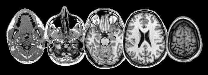
CT Scan on the left, MRI on the right.  You see how the bone on these scans don't match up?
That screams ERROR to me.

     You see, millimeter accuracy is needed for successful DBS surgery largely because of the small STN size, but even more importantly because the STN is close (1-3mm) to the anatomic structures such as the red nucleus, posterior limb of the internal capsule, medial lemniscus, and substantia nigra, the stimulation of which may result in adverse reactions.   
     The only good thing to come out of this mess is that MRI distortions occur most significantly on the edges of the image, and the middle (where these structures are) are less likely to be subject to significant distortion.   

Image Fusing: Pre-op and post op scans must be fused together in order to properly estimate errors in electrode placement.  Image fusing is a frustrating thing to do because in order for some parts of the image to line up other parts will not.  And that just creates (you guessed it) more error.  This error can present itself also in millimeter amounts, but measurement estimations can differ from person to person.  

Brain Shift: You can't control this one.  Brain shift occurs naturally because your brain is floating around in a hard shell (skull) so naturally if you tilt your head, it will move slightly toward the direction of gravity.  I wrote two rather long articles on brain shifts (which you can read here and here) so I won't elaborate much on it now.  What I will say is one way to avoid the brain shifting too much is imaging the patient in the same position as the surgery will be done, so at least the brain won't shift due to patient movement.  
     Some DBS centers have tried to drain all the CSF out of the brain before doing the operation and imaging so no brain shift can occur (theoretically).  The brain sinks to the bottom of the skull during the operation, but once the CSF fills the skull again (and the brain returns to its original position) the electrode will shift in the brain (another error).

In general, there is nothing we can do to avoid these errors (these aren't all the errors either).  I sincerely hope reading this post hasn't resulting in any stressful hair loss. I'm sad to report the same cannot be applied to me.


More on errors in a future post!


Holly

Week 6: It's All In the Details + Musings From a Student on a Stool Gazing Longingly into the OR

Greetings again!

   I arrived at the hospital at 6:30 am yesterday morning ready to observe a Stage I DBS electrode placement operation.  The residents were already bustling about when I walked into the pre-op rooms (apparently everyone is a morning person here).  I was able to witness Dr. Kasoff attach a Leksell frame to a patient's head, which had a sad undertone to it.  The patient's head was numbed under local anesthesia and the frame is attached via screws.  Securing the metal rectangle looks extremely uncomfortable, and by the expression on the patient's face, he wasn't having the best time either.


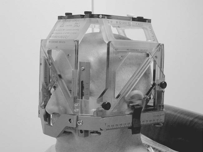
     The half circle apparatus isn't attached until the patient is in the OR.  What the picture isn't showing is this plastic cage (the localizer) that is attached to the frame when the patient gets a pre-op CT scan.


This (localizer) is attached to the Leksell frame when the patient gets a CT scan.  This localizer provides axises for the surgeon to pinpoint his target. 


A different localizer is used, but this is the general idea.
Who knew it was so difficult to find images of extremely specific relatively new neurosurgery apparatuses?

After the pre-op CT scan, the patient is wheeled into the OR and the operation begins.  Because I was not in the room during the procedure, I can't talk much on the details, but here is what I gathered:


1. The patient is 'asleep' at the beginning and end of the operation.  That is, he/she is unconscious when the surgeon is making the incision, drilling the hole, and opening the dura (and closing).  It makes a lot of sense because I think it would be extremely difficult to remain calm if I heard someone drilling into my skull.  There is most likely a more scientific explanation to why the patient is 
unconscious and I will update this post if I out the reason.

2. The patient has to be awake when the electrode is being inserted to ensure that a desired clinical outcome is achieved.  This ties into the whole debate of anatomy vs. physiology that I will expand on in a later post.  If the tremor is still present, the surgeon often will do multiple runs with the electrode. 


3. If the tremor is temporarily suppressed right after the electrode is inserted (without any electrical stimulation), that's a pretty good indication that the electrode is in a good spot.


4. Sometimes the patient will feel tingling when there is a current.  Sometimes the tingling will disappear within a minute or two (this is good).  If the tingling persists, the electrode needs to be adjusted because it is uncomfortable to the patient.  Where the tingling is located gives the surgeon a pretty good idea how to adjust the electrode. 


5. In addition to checking for tremor suppression, the doctors also check for slurred speech (or lack thereof) and the breadth of the visual field.  The patients are asked to count backwards from 10, drink from an empty cup (no fluids before or during surgery!), and touch their finger to a doctor's moving hand.


6. After the electrode is inserted, the patient is asked to move around certain parts of his body (or have the doctors poke and prod at his body).  If the electrode is in the correct position, you can actually hear these movements via neural noise.  Neural noise is this static sound.  For example, if you move your jaw, the static sound may start popping at a high frequency.  


That's all I have to today.  I wasn't even in the operating room and I was a bit exhausted trying to keep up with all the technical details (from the research papers).  The entire operation was close to eight hours long.  More posts are coming up about the setup of this project and some more background information that I still need to mention.


Phew! I hope I will be able to build up endurance by watching more and more of these surgeries (and not internally cry every time we take the stairs from the first floor surgery rooms to the offices on the fourth floor).  My calf muscles have really been shaping up while I stand by the window of the OR.  Compared to the ever chirpy doctors and nurses, I still have a long way to go (and many more cups of coffee to drink).


Holly


Week 5: The Chronicles of a High School Student Observing a Stage II DBS Surgery

Warmest Greetings!

*********One graphic image************
     This may be a long post, so pour yourself a cup of tea and grab a biscuit (or not if you're queasy) before settling in.
     I have been repeatedly told that the Stage II DBS surgery is the "unglamourous" side of neurosurgery and employs the mentality of "what needs to be done needs to be done".  You will see what I mean very quickly.
     Stage II DBS surgery consists of the surgeon connecting a wire from the electrodes implanted in the brain to an IPG (implanted pulse generator - basically a battery pack) surgically inserted into the clavicle region.

This is an example of an IPG.

This is roughly where an IPG is implanted.
     The operation seems to be quite straightforward: the surgeon makes an incision and a small pocket in the upper chest/clavicle region for the IPG to sit and a wire is strung underneath the skin connecting said IPG to the electrodes on the skull (previously placed in the Stage I operation).

.....but how is the wire "strung"?

     A metal tube (diameter of roughly 5mm) called the tunneling rod is manually pushed under the patient's skin.  Yes.  Manually pushed.  I was debating whether or not I was going to get light headed watching two surgeons trying to push the tube across a length of the patient's neck.  The scrub nurse admitted that the first time she was in on the operation, she couldn't watch.

An example of what a tunneling tool looks like.
     It's no surprise that this wire is what patients complain mostly about.  A representative from the Medtronics company said that patients will often feel pain or soreness in the area around where the wire is inserted.  Apparently, not many nerves or blood vessels are damaged.  If there is bleeding (more than normal), once pressure is applied, it will stop.
     Otherwise, watching this operation was a pretty pleasant experience.  Everyone was extremely accommodating and understanding.  There are many steps before and after the procedure that medical shows like Grey's Anatomy or Hopkins Med cut out.  Everyone wears scrubs because they are easy to clean and easy to replace.  I wear paper scrubs because I'm an observer.  No outside clothing can be exposed in the operating room (and the rooms around it) so shoe covers and hair caps are mandatory.  A face mask must be worn at all times in the OR.
Neurosurgeons also wear funny magnifying glasses and a head light.  

Fierce
This is what I look like, except with shoe caps and my shirt tucked in cause I'm professional.

     A scrub nurse will help gown and glove the surgeon and any assisting residents.  Anyone who handles the sterile medical equipment wears two gloves after scrubbing in.  Keeping everything sterile is so important that some medical equipment is doubled packaged.  There is a red square in the center of the room where the patient and surgeons are, and it is dubbed (in my head) the sterility square.


     I watch from outside because I am not of age yet.  Before the operation starts, everyone in the OR states their name and their role.  The surgeon is the top dog in the OR.  Whatever he/she needs, there will be at least three pairs of hands helping him efficiently perform the operation.  The scrub nurse maintains the blades, sutures, irrigation, etc.  There is periodic equipment counting to ensure that all objects are in their rightful places.  The gauze, needles, blades etc are all tallied on the board to double check that nothing is left in the patient.
     Blades are numbered by their size.  The bigger the number the smaller the blade.  The same concept goes for needles.  The two scalpels used in this operation were the 10 blade and 15 blade.


     After the surgery, the surgeon records and dictates the events of the surgery and calls up the family.

So.....what's it like observing an operation?  Really intriguing.  Kinda chilly.  Completely awestruck.  One perk of doing my project in the hospital: everyone is really kind.  People go into the medical field because they want to help others, so the work environment is cheery and jolly (and occasionally sassy).
     When the patient was wheeled out and the room was cleaned, I realized how sore my body was standing in one position for hours (and I wasn't even operating!).  On top of that, doctors walk fast.  Just in general.  They stride efficiently with purpose.  I scamper along after them.
     Needless to say, I now have buckets of respect (even more than before) for the nurses, surgeons, residents, and hospital staff in general for working long shifts and always being on top of their game.  After being thoroughly amazed by how to human body can withstand surgical procedures and re-motivated to hit the books, the main thought as I get into the car to drive home is:


Holly


Image Sources:
http://www.implantable-device.com/2013/12/05/medtronic-implants-activa-pcs-dbs-ipg-in-us/
http://www.tedpella.com/dissect_html/549-3-blades.jpg
http://parkinsonshumor.blogspot.com/2012/11/parkinsons-dbs-surgery-part-4-day.html
http://www.basicspine.com/blog/wp-content/uploads/surgeon-operating.jpg?8212ac


Week 4: Parkinson's Disease 101

Hello again!

     I guess in my haste trying to introduce this project, I failed to mention the cardinal traits of Parkinson's disease.  When shadowing Dr. Kasoff, I was able to see first hand a few of these PD symptoms.  


A patient presenting with some symptoms of Parkinson's Disease (PD).


1.) Resting tremor: This cardinal trait is what we widely associate with Parkinson's Disease patients.  Patients present with an involuntary shaking in their arms and/or legs (it's uncommon for tremors to occur in the jaw, face, or trunk, but still possible) when the muscles are relaxed, hence the term "resting tremor".  The resting tremor can be emphasized when the patient is distracted.  In other words, sometimes the doctor will ask the patient to perform simple arithmetic problems in their head so the resting tremor can be "brought out" more (Ms. K you were right; mental math is always lurking around the corner).  The tremor usually starts in one finger then progresses to the hand etc before passing to the other side.  If the tremor is only present on one side of the body, then only one hemisphere of the brain will be implanted with an electrode (unilateral DBS, one burr hole).  And if the patient experiences tremors on both sides, then both sides of the brain will be stimulated (bilateral DBS, two burr holes).


2.) Rigid movement: The term "cog-wheel rigidity" is often coined to describe the motion of Parkinson's patients.  Similar to how a cog-wheel spins, the movement of a PD patient is a series of frequent pauses and jerky quick movements.  The muscle tone of PD patients is always stiff which reduces their range of motion (ex. not swinging arms when walking).  As you can imagine, this is extremely uncomfortable and in some cases can be painful.


3.) Bradykinesia: This is a fancy term for slow movement, but can also mean a general reduction of spontaneous movement.  Patients presenting with bradykinesia are often abnormally still and struggle with repetitive movements like brushing their teeth, tapping their fingers, and walking (they walk in short shuffling steps).  


4.) Postural Instability: Postural instability is exactly what it sounds like: the tendency for a patient to lose balance when standing, getting up, turning, or being pushed.  I realize that pushing a PD patient sounds absolutely cruel, but in order to test for postural instability, doctors will pull (with some force) the patient and see how he/she recovers.  A person without PD will adjust his footing when pulled but a PD patient will topple backwards (safely into the arms of the doctor).


     Those are the four cardinal traits of Parkinson's Disease, but there are many other symptoms you can look for.  The list is quite long, so I will only mention the two that I saw.


Hypophonia: soft speech can be a result of bradykinesia

Hypomimia: masked face (little to no facial expressions) can also be a result of bradykinesia

     Now you must be wondering, how do you get Parkinson's Disease?  We don't know exactly.  There is a very very small genetic component, but we do know that patients with PD have fewer dopamine cells.


     That's all for now.  Stay tuned for some surgery experiences in the next post!

Holly


Image Source: http://www.doctortipster.com/10064-parkinson-disease-symptoms-and-diagnosis.html
http://blogs.brandeis.edu/flyonthewall/files/2014/08/Post4_1.jpeg
Sources: http://www.pdf.org/symptoms_primary